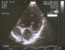
Ultrasonography uses high-frequency sound waves to produce images of internal organs and other structures inside the body. It can be used to assess the animal’s heart, kidneys, liver, and bladder. It can help detect fluid, cysts, tumors or abscesses, and also to confirm pregnancy or to monitor an ongoing pregnancy. This imaging technique may be used in combination with radiographs and other diagnostic methods to ensure a proper diagnosis.
A small hand-held tool called a transducer is applied to the surface of the body to record images of the area of interest. Gel is used to help the transducer slide over the skin surface and create a more accurate visual image.
Sound waves are emitted from the transducer and are directed toward the structures to be examined. The sound waves create echoes of various degrees which depend on how dense the tissue is and the amount of fluid that is present. The waves create thorough images which are shown on a viewing monitor and recorded for evaluation.
Ultrasound is a painless procedure with no known side effects and does not involve radiation. This procedure does not typically require pets to be sedated or anesthetized.
If you have questions about our Ultrasonography services, please don’t hesitate to ask.